
With ultrasound we can assess the pleura and the areas of the lung close to the surface directly in our practice, without radiation and with an immediate result.
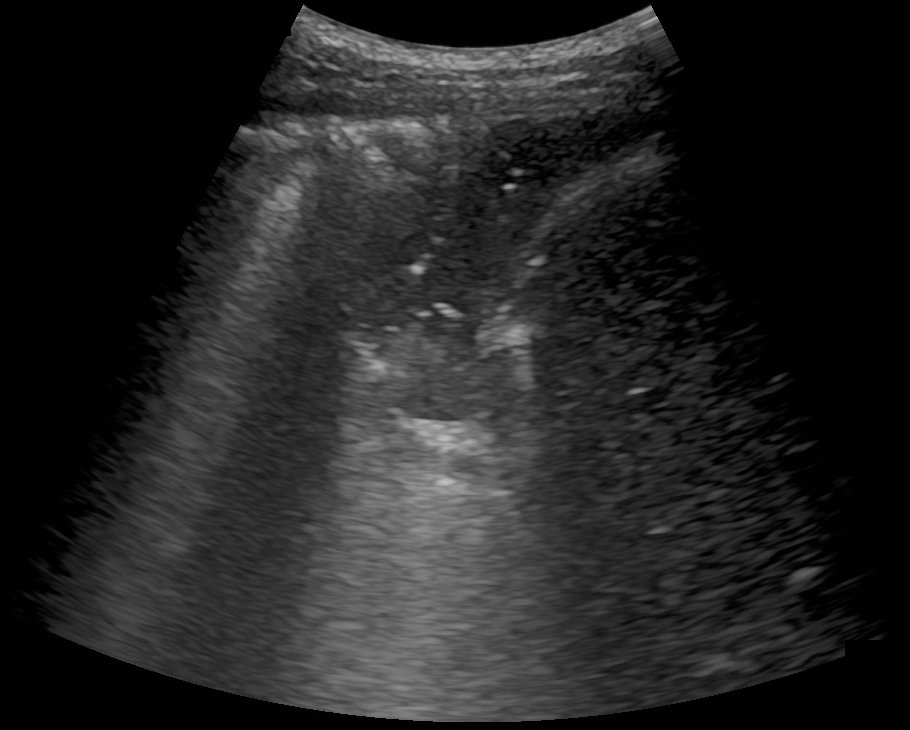
When do we use thoracic ultrasound?
- if fluid in the chest (pleural effusion) is suspected
- if pneumonia is suspected, and to monitor the course of treatment
- for chest pain that depends on breathing, e.g. if pleurisy is suspected
- for shortness of breath, for example to detect fluid in the lungs due to heart failure
- if a collapsed lung (pneumothorax) is suspected
Advantages
The examination puts no strain on you and can be repeated as often as needed. This makes it particularly suitable for follow-up checks and for patients in whom X-rays should be avoided where possible, for example during pregnancy.
Limitations
Changes deep inside the lung that do not reach the surface cannot be seen on ultrasound. In these cases we arrange an X-ray or a CT scan if necessary.
Procedure
You usually sit upright with your upper body undressed. We examine the chest, sides and back using a little ultrasound gel. The examination takes about 10 to 15 minutes, and no preparation is needed. We discuss the result with you straight away.